
Tissue
· Tissue is the groups of cells which perform specialized activity of the body.
· Tissue is the group of cells, which are similar in origin, structure and function are called as “Tissue”.
· The study of tissue is called “Histology”.
· The tissue in plants is divided into three types;
o Vascular tissue
o Ground tissue
o Epidermal tissue
· Body tissues can be classified into four basic types according to their structure and function.
o Epithelial tissue
o Connective tissue
o Muscular tissue
o Nervous tissue
Epithelial tissue
· This type of tissue covers the entire body and lines cavities, hollow organs, and tubes. It also is found in glands.
· Function
o Protection from dehydration, chemical and mechanical damage
o Secretion
o Absorption
· The cells are closely packed, and the intercellular material, known as the matrix.
· The cells often lie on a basement membrane, which is an inert connective tissue formed by the epithelial cells themselves.
· Epithelial tissue divided into
o Simple epithelium (single layer of cells)
o Stratified epithelium (several layers of cells)
Simple epithelium
· Simple epithelium consists of a single layer of identical cells and is classified into three categories.
o Squamous epithelium
o Cuboidal epithelium
Columnar epithelium
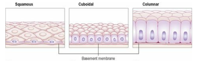
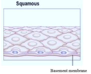
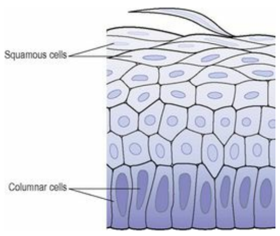
Squamous epithelium
· This is composed of a single layer of flattened cells.
· The cells are arranged on top of each other like flat stones, creating a very thin and smooth membrane that diffuses easily.
· Location:
o heart, blood vessels, lymphatic vessel linings – known as endocardium
o peritoneum, pleura, pericardium – called mesothelium
o Alveoli of the lungs
o Lining the collecting ducts of nephrons in the kidneys.
· Function
o Blood filtration in kidneys
o Diffusion of oxygen into blood vessels of lungs
o Site of secretion in serous membranes
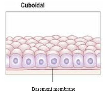
Cuboidal epithelium
· This consists of cube-shaped cells fitting closely together lying on a basement membrane.
· Function
o It forms the kidney tubules and is also present in some glands.
o Lies anterior surface of capsule of lens and forms pigmented epithelium at posterior surface of retina of the eye
· Function
o Cuboidal epithelium is actively involved in secretion, absorption and excretion.
Columnar epithelia
· This is formed by a single layer of cells, rectangular in shape, on a basement membrane.
· It lies many organs and possesses to a specific function;
· The lining of the stomach is formed from simple columnar epithelium without surface structures.
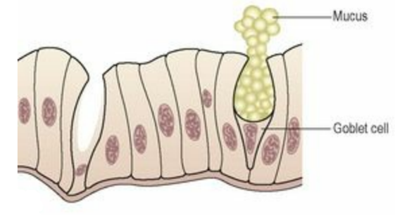
· The surface of the columnar epithelium lining the small intestine is covered with microvilli.
· Microvilli have a wide surface area for absorption of nutrients from the small intestine. The trachea’s columnar epithelium is ciliated and also contains goblet cells that secrete mucus.
Pseudostratified columnar epithelium
· All cells are attached to basement membrane in a single layer, but some cells do not extend to apical surface.
· Pseudostratified ciliated columnar epithelium contains cells that extend to surface and secrete mucus (goblet cells) or bear cilia.
· Pseudostratified non-ciliated columnar epithelium contains cells without cilia and lacks goblet cells.
· Location
o Upper respiratory tract; non-ciliated variety lines larger ducts of many glands, epididymis, and part of male urethra.
· Function
o Ciliated variety secretes mucus that traps foreign particles, and cilia sweep away mucus for elimination from body; non-ciliated variety functions in absorption and protection.
Stratified epithelia
· Stratified epithelia are made up of many layers of cells of various shapes.
· Basement membranes are usually absent.
· The primary function of stratified epithelium is to protect the underlying tissues from mechanical wear and tear.
· There are two main types:
1. Stratified squamous epithelium
2. Transitional epithelium
Stratified squamous epithelium
· The stratified squamous epithelium is a type of tissue found covering and lining parts of the body.
· In this tissue, cells are flattened, joined tightly together, and stacked.
it is found in the outermost layer of the skin and the lining of the esophagus, mouth and female reproductive organ.
Keratinised stratified epithelium
· This is present on dry surfaces like skin, hair, and nails.
· The surface layer is composed of dead epithelial cells that have lost their nuclei and contain the protein keratin.
· This creates a protective coating that is strong and somewhat waterproof, preventing the living cells underneath from drying out.
Non-keratinised stratified epithelium
· This preserves wet surfaces prone to wear and strain and prevents them from drying out, such as the conjunctiva of the eyes, the lining of the mouth, the throat, the esophagus, and the vagina.
Function
· Protection against abrasion, water loss, ultraviolet radiation, and foreign invasion. Both types form first line of defense against microbes.
Stratified cuboidal epithelium
· It is a rare type of epithelia contain cuboidal shaped cells arranged in multiple layers.
· Location
o Ducts of adult sweat glands and oesophageal glands, part of male urethra.
· Function
o Protection; limited secretion and absorption.
Stratified columnar epithelium
· It is usually consisting of shortened, irregularly shaped cells.
· Location
o Lines part of urethra, esophageal glands, anal mucous membrane, and part of conjunctiva of eye.
· Function
o Protection and secretion.
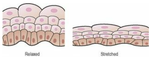
Transitional epithelium
· This consists of multiple layers of pear-shaped cells. It lines the urine bladder and allows it to extend when it fills.
· Location
o Lines urinary bladder and portions of ureters and urethra.
· Function
o Allows urinary organs to stretch and maintain protective lining while holding variable amounts of fluid without rupturing.
Connective tissue
· Connective tissue is one of the most abundant and widely distributed tissues in the body.
· Function
o It binds together, supports, and strengthens other body tissues
o Protects and insulates internal organs
o It distinguishes structures such as skeletal muscles
o Serves as the major transport system within the body (blood, a fluid connective tissue)
o Primary location of stored energy reserves (adipose, or fat, tissue)
o The main source of immune responses
Connective Tissue Cells
1. Fibroblast
· It is flat cells with branching processes. They are present in all the general connective tissues, and usually are the most numerous.
· Fibroblasts migrate through the connective tissues, secreting the fibers and certain components of the ground substance of the extracellular matrix.
2. Macrophages
· Macrophages develop from monocytes, a type of white blood cell. Macrophages have an irregular shape with short branching projections and are capable of engulfing bacteria and cellular debris by phagocytosis.
3. Plasma cells
· These develop from B-lymphocytes, a type of white blood cell.
· They synthesise and secrete specific defensive antibodies into the blood and tissues.
4. Mast cell
· They are found in loose connective tissue and under the fibrous capsule of some organs, e.g. liver and spleen, and in considerable numbers round blood vessels.
· They produce granules containing heparin, histamine and other substances, which are released when the cells are damaged by disease or injury.
· Histamine is involved in local and general inflammatory reactions; it stimulates the secretion of gastric juice and is associated with the development of allergies and hypersensitivity states.
· Heparin prevents coagulation of blood, which may aid the passage of protective substances from blood to affected tissues.
5. Adipocytes
· Adipose cells, are connective tissue cells that store triglycerides (fats).
· They are found deep to the skin and around organs such as the heart and kidneys.
6. Leukocytes (White blood cells)
· White blood cells are normally found in small numbers in healthy connective tissue but neutrophils migrate in significant numbers during infection when they play an important part in tissue defence.
Connective Tissue Extracellular Matrix
· The extracellular matrix consists of two major components
1. The ground substance and
2. The fibers.
Ground substance
· The ground substance is the component of a connective tissue between the cells and fibers.
· It may be fluid, semifluid, or gelatinous.
· Function
o Supports cells and binds them together
o Store water
o Exchange of substances between the blood and cells
o Active role in tissue development, migrate, proliferation, and shape change
o Metabolic function
· Ground substances also contain complex combinations of polysaccharides (hyaluronic acid, chondroitin sulfate, dermatan sulfate, and keratan sulfate) and proteins.
Fibres
· Three types of fibers are embedded in the extracellular matrix between the cells:
1. Collagen fibers
2. Elastic fibers
3. Reticular fibers
Collagen fibers
· Collagen fibres are fibrous proteins and are secreted into the extracellular space and they provide high tensile strength to the matrix.
· Mostly found in cartilage, bones, tendons, ligaments, and skin.
Elastic fibres
· Elastic fibres are long, thin fibres that form branching network in the extracellular matrix. They help the connective tissue to stretch and recoil.
· Present in skin, blood vessel walls, and lung tissue.
Reticular fibers
· Reticular fibers are short, fine collagenous fibres that can branch extensively to form a delicate network.
· Found in spleen and lymph nodes.
Classification of Connective Tissue
1) Embryonic connective tissue
a) Mesenchyme
b) Mucous connective tissue
2) Mature connective tissue
a) Loose connective tissue
i) Areolar connective tissue
ii) Adipose tissue
iii) Reticular connective tissue
b) Dense connective tissue
i) Dense regular connective tissue
ii) Dense irregular connective tissue
iii) Elastic connective tissue
c) Cartilage
i) Hyaline cartilage
ii) Fibrocartilage
iii) Elastic cartilage
d) Bone tissue
e) Liquid connective tissue
i) Blood tissue
ii) Lymph
Embryonic Connective Tissue
· Embryonic connective tissue is of two types: mesenchyme and mucous connective tissue.
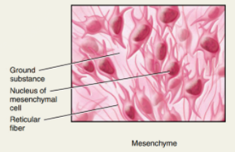
Mesenchyme
· It has irregularly shaped mesenchymal cells embedded in semifluid ground substance that contains delicate reticular fibers.
· Location
o Almost exclusively under skin and along developing bones of embryo; some in adult connective tissue, especially along blood vessels.
· Function
o Forms almost all other types of connective tissue
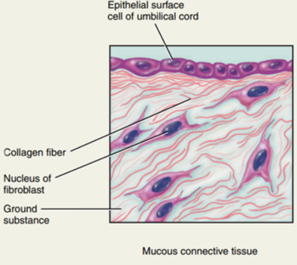
Mucous connective tissue
· Mucous connective tissue has widely scattered fibroblasts embedded in viscous, jellylike ground substance that contains fi ne collagen fibers.
· Location
o Umbilical cord of foetus.
· Function
o Support
Mature Connective Tissue: Loose Connective Tissue
· Mature connective tissue, is present in the newborn. Its cells arise primarily from mesenchyme.
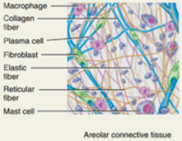
Areolar connective tissue
· Areolar connective tissue is one of the most widely distributed connective tissues; consists of fibers (collagen, elastic, reticular) arranged randomly and several kinds of cells (fibroblasts, macrophages, plasma cells, adipocytes, mast cells, and a few white blood cells) embedded in semifluid ground substance (hyaluronic acid, chondroitin sulfate, dermatan sulfate, and keratan sulfate).
· Location
o In and around nearly every body structure, subcutaneous layer deep to skin papillary region of dermis of skin, lamina propria of mucous membranes, around blood vessels, nerves, and body organs.
· Function
o Strength, elasticity, support.
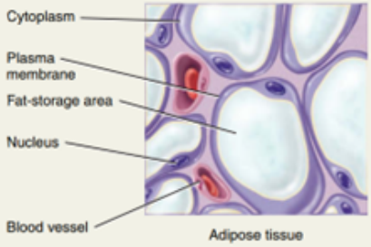
Adipose tissue
· Adipose tissue has cells derived from fibroblasts (called adipocytes) that are specialized for storage of triglycerides (fats), centrally located droplet. Cell fills up with a single, large triglyceride droplet, and cytoplasm and nucleus are pushed to periphery of cell.
· Location
o subcutaneous layer deep to skin, around heart and kidneys, yellow bone marrow, padding around joints and behind eyeball in eye socket
· function
o Reduces heat loss through skin; serves as an energy reserve; supports and protects organs. In newborns, BAT generates heat to maintain proper body temperature.
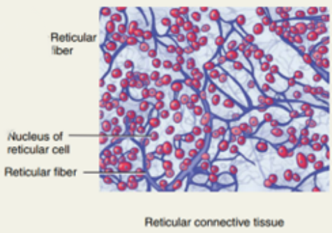
Reticular connective tissue
· Reticular connective tissue is a fi ne interlacing network of reticular fibers (thin form of collagen fibre) and reticular cells.
· Location
o Stroma (supporting framework) of liver, spleen, lymph nodes; red bone marrow; reticular lamina of basement membrane; around blood vessels and muscles.
· Function
o Forms stroma of organs; binds smooth muscle tissue cells; filters and removes worn-out blood cells in spleen and microbes in lymph nodes.
Dense connective tissue
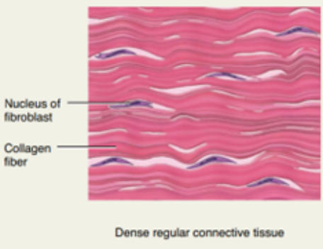
Dense regular connective tissue
· Dense regular connective tissue forms shiny white extracellular matrix; mainly collagen fibers regularly arranged in bundles with fibroblasts in rows between them. Collagen fibers (protein structures secreted by fibroblasts) are not living, so damaged tendons and ligaments heal slowly.
· Location
o Forms tendons (attach muscle to bone), most ligaments (attach bone to bone), and aponeuroses (sheetlike tendons that attach muscle to muscle or muscle to bone).
· Function
o Provides strong attachment between various structures. Tissue structure withstands pulling (tension) along long axis of fibers.
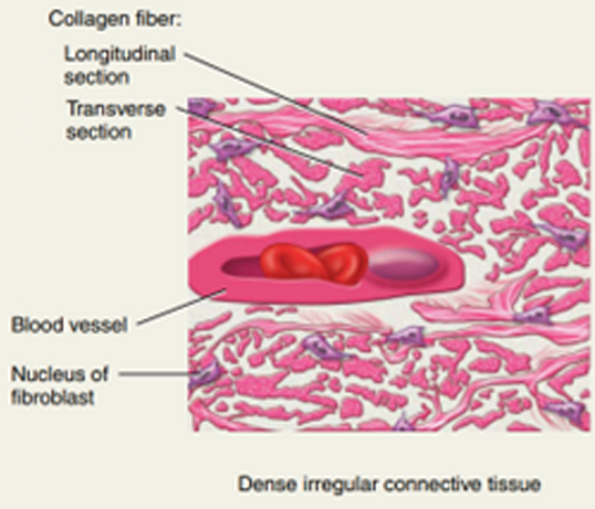
Dense irregular connective tissue
· Dense irregular connective tissue is made up of collagen fibers; usually irregularly arranged with a few fibroblasts.
· Location
o tissue beneath skin and around muscles and other organs, fibrous pericardium of heart, periosteum of bone, perichondrium of cartilage, joint capsules, membrane capsules around various organs (kidneys, liver, testes, lymph nodes); also, in heart valves.
· Function
o Provides tensile (pulling) strength in many directions.
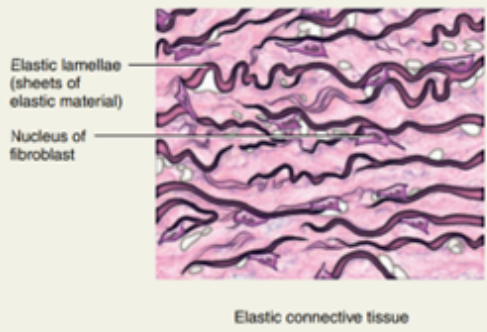
Elastic connective tissue
· Elastic connective tissue contains predominantly elastic fibers with fibroblasts between them; unstained tissue is yellowish.
· Location
o Lung tissue, walls of elastic arteries, trachea, bronchial tubes, true vocal cords, suspensory ligaments of penis, some ligaments between vertebrae
· Function
o Allows stretching of various organs; is strong and can recoil to original shape after being stretched. Elasticity is important to normal functioning of lung tissue (recoils in exhaling) and elastic arteries (recoil between heartbeats to help maintain blood flow).
Cartilage
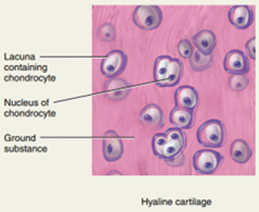
Hyaline cartilage
· Hyaline cartilage contains a resilient gel as ground substance and appears in the body as a bluish-white, shiny substance; prominent chondrocytes are found in lacunae surrounded by perichondrium (exceptions: articular cartilage in joints and cartilage of epiphyseal plates, where bones lengthen during growth).
· Location
o Most abundant cartilage in body; at ends of long bones, anterior ends of ribs, nose, parts of larynx, trachea, bronchi, bronchial tubes, embryonic and fetal skeleton.
· Function
Provides smooth surfaces for movement at joints, flexibility, and support; weakest type of cartilage and can be fractured.
Fibrocartilage
· Fibrocartilage has chondrocytes among clearly visible thick bundles of collagen fibers within extracellular matrix; lacks perichondrium.
· Location
o Pubic symphysis (where hip bones join anteriorly), intervertebral discs, menisci (cartilage pads) of knee, portions of tendons that insert into cartilage.
· Function
o Support and joining structures together. Strength and rigidity make it the strongest type of cartilage.

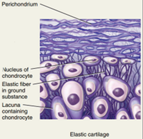
Elastic cartilage
· Elastic cartilage has chondrocytes in threadlike network of elastic fibers within extracellular matrix; perichondrium present.
· Location
o Lid on top of larynx (epiglottis), part of external ear (auricle), auditory (eustachian) tubes.
· Function
o Provides strength and elasticity; maintains shape of certain structures.

Hi….!! My name is Smrutiranjan Dash, From Odisha, India. Professionally I am Assistant Professor at The Pharmaceutical College, Barpali, Odisha, department of Pharmacology.