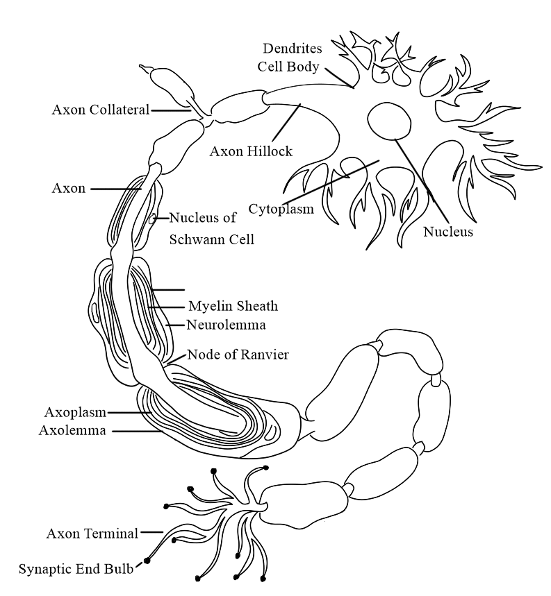
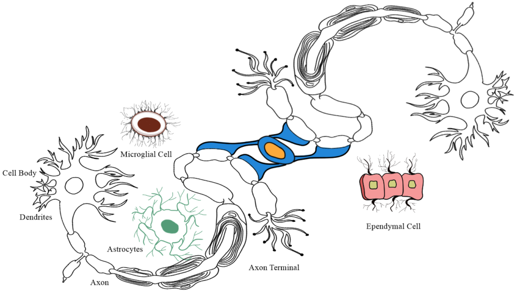
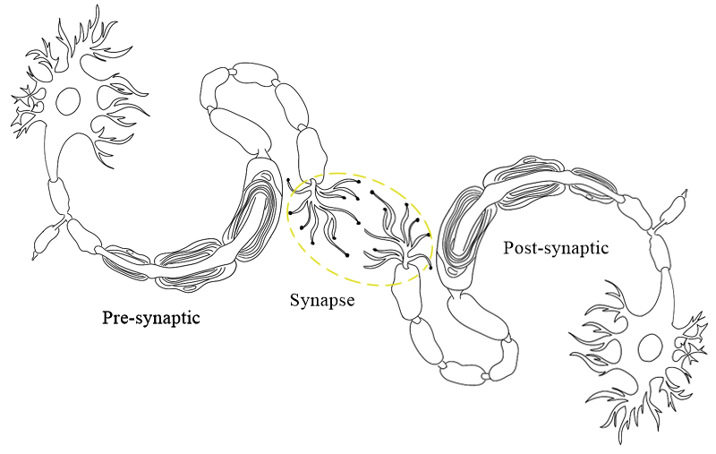
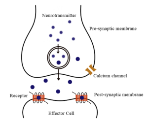
THE NERVOUS SYSTEM

THE NERVOUS SYSTEM

·         The nervous system detects and responds to change inside and outside the body.

·         It controls many vital aspects of body function and maintain homeostasis.

 ·         The nervous system has three main function.

1.      Sensory

2.      Integration

3.      Motor 

Classification 

·         The nervous system consists of neurons, which conduct nerve impulses and are supported by unique connective tissue cells known as Neuroglia.

Sensory: The chemical messenger relay into the brain.

Integration: To analyse and process the sensory input.

Motor: To respond the message to appropriate organs.

Somatic nervous system (Voluntary): This system consists of sensory neurons that conducts
impulse from somatic and special sense receptors to the CNS and motor neurons
from the CNS to skeletal muscle.

Autonomic nervous system
(Involuntary):
It contains sensory
neurons from motor neurons that convey impulses from CNS to skeletal muscle
tissue or different periphery and glands.

Example: Breath, Digestion, Heart rate etc.

Nervous system consists of two specific type of cells;

           1.      Neurons

     2.    Neuroglia 

NEURONS

·         Neurons are the fundamental units of the brain and nervous system, the cells that receive and sends message from the body to brain and back to the body.

·         Each neuron consists of a cell body and its processes one axon and many dendrites.

·         Neurons need continuous supply of O2 and glucose for their survival.

Axon and dendrites

·         Axon and dendrites are extensions of cell bodies.

Axon:

·         Each nerve cell has only one axon, which begins at a lappered area of the cell body, that is called Axon Hillock.

·         It carries impulses away from the cell body and are usually longer than the dendrites sometimes it is about 100cm long).

·         The membrane of the axon is called Axolemma.

·         Large axons and those of peripheral nerves are surrounded by a myelin sheath.

·         The axon in covered by number of layers of Schwann cell plasma membrane.

·         Between the layers of plasma membrane there is small amount fatty substance called myelin.

·         The outer most layer of Schwann cell plasma membrane is the Neurilemma.

Dendrites

·         It receives and carry impulses towards cell bodies.

·         They have the same structure as axon but are usually shorter and branching.

·         In motor neurons dendrites form part of synapse and in sensory neurons they form the sensory receptors that respond to specific stimuli.

Neuroglia cells

·         It is non-neuronal cells (doesn’t make neurons structure)

·         Neuroglia cells provides mechanical and metabolic support to the neurons.


·         These cells perform support roles for the neurons to ensure their survival.

In CNS

Astrocytes cells

·         It is a star like appearance so that it called astrocytes.

·         The cell body usually attached between blood capillary and neuron of the brain.

·         The astrocytes regulate flow of ions, sugar, oxygen and carbon dioxide into and out of the neurons.

·         Astrocytes prevents toxic substances from entering brain neurons.

Ependymal cell

·         The ependymal cell usually cubed shaped.

·         These cells are ciliated-epithelial glial cells lining the spinal cord and cerebral ventricles.

·         It produces cerebrospinal fluid.

·         It protects the brain and spinal cord both mechanically and immunologically.

·         Ependymal cell contains cilia that helps to circulate cerebrospinal fluid.

Microglial cells

·         Microglial cell is a smallest neuroglial cell and able to migrate around the CNS.

·         It supports neuron by phagocytosis of bacterial cells and dead cell debris.

Oligodendrocytes cells

·         These are the myelinating cells of the central nervous system.

·         They provide insulating layers of the myelin around the axon within the brain and spinal cord.

In PNS

Schwann cells

·         The PNS contain neuroglial cell as well as Schwann cells.

·         It forms a covering called myelin sheath around the axons.

·         They perform the same role as the oligodendrocytes found in the CNS.

Satellite cell

·         These are the small glial cell that surround neurons sensory ganglia in the ANS.

·         PNS satellite glia cells are very sensitive to injury and exacerbate pathological pain.

·         It gives protection to the neuron cells.

Electrophysiology

·         it includes measurement of the electrical activity of the neurons or action potential.


·         It indicates the flow of ions in biological tissues.

·         The cell membrane of every cell contains ion that are present inside and outside of the membrane.

·         Na+ ion have high concentration at outside the cell so it is called extracellular ion.

·         K+ ion have high concentration at inside the cell so it is called intracellular ion.

·         At resting state: The ions are flows by the concentration gradient (Higher to lower) and becomes polarized.

·         Due to change in concentration of ion at inside and outside the cells it develops potential difference.

·         At resting state, it is -70mv (Resting potential).

Threshold potential

·         When a stimulus arises in neurons it changes the resting membrane potential and it became depolarization.

·         Example, temperature, pressure, chemical etc.

·         The stimulus changes resting potential (-70mv) to -55mv then it is called threshold potential.

Action potential

·         When the stimulus achieved at threshold potential it open Na+ channel. Due to high concentration of Na+ ions are entering inside the cell. There are change f chare become positive that is called depolarization.

·         At the same time K+ channel also open and outflux of K+ ions. Again, change charged to (-ve). It is called repolarization.

Threshold potential

Na+ channel open

Influx of Na+ ion

DEPOLARIZATION

At the same time

K+ channel open

Outflux of K+ ion

REPOLARIZATION


Nerve impulse

·         It is a signal that transmitted along a nerve fibre.

·         The information or stimulus that is transfer through the neurons.

·         It transmitted through axon by propagation of action potential.

Receptor

·         Receptors are the macro molecules made up with proteins, which are those binding sites where the neurotransmitter binds.

·         Neurotransmitter receptor are present in post synaptic membrane.

·         There are two types of neurotransmitter receptors

                                i.            Ionotropic receptor

                              ii.            Metabotropic receptor


Ionotropic receptor (Ligand-gated-ion-channel)


·         When neurotransmitter attached to their site it open or close ion channel according to NT nature.

Metabotropic receptor (G Protein Coupled Receptor)


·         When neurotransmitter attached with receptors will open or close ion channel or direct or by indirectly (through secondary messenger)

Synapse


·         The junctions of two neurons where the information is passed to the next.

Types of synapse

1.      Electrical synapse (faster transmission)

2.      Chemical synapse

Electrical synapse

·         In an electrical synapse the axon of one neuron is connected to dendrites of another neuron by gaps.

·         The gap junction is composed of channel protein known as connexins.


·         Connexins constitutes a large family of trans membrane proteins that allow intracellular communication and the transfer of ions and small signaling molecules between cells.

Chemical synapse

·         Chemical synapse release of Chemical NTs to propagate signal from one neuron(pre) to another (post synaptic).

·         The distance between two neurons is about 20-30nm called synaptic cleft.

·         At the end of the neuron, is axon terminal there are present synaptic vesicles contains NT.

·         So, when AP reaches at nerve terminal the vesicles are ruptured and the NTs are released to post synaptic membrane.                
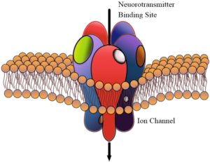
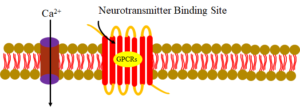
 Neurotransmitter

  • A neurotransmitter is a signalling molecule released by neuron to affect another neuron across a synapse.

Types of neurotransmitter

Excitatory

Inhibitory

Both

·         Glutamate

·         Aspartate

·         GABA

·         Serotonin

·         Dopamine

·         Acetylcholine

·         Nor-adrenalin

 Adrenaline

  • Adrenaline is a primary hormone released by the adrenal gland, but some neurons may secrete it as a neurotransmitter.
  • Increased heart rate and blood flow.
  • Produces during stressful or exciting situation.

Dopamine

  • It is primarily responsible for feeling of pleasures.

Acetyl Choline

  • Involved in thought, learning and memory within the brain.
  • Activates muscle contraction.

Serotonin


  • It is an inhibitory neurotransmitter that regulates mood, fear, feeling of relaxation, mental focus.
  • Regulates Digestion, nutrition absorption.

Nerves

·         It consists of numerous neurons collected into bundles. Each bundle has several coverings of protective connective tissue.

o   Endoneurium

o   Perineurium

o   Epineurium

Sensory or afferent nerves

·         Sensory nerves carry information from the body to spinal cord.

·         The impulses may then pass to the brain or to connector neurons of reflex arcs in the spinal cord.

Sensory receptor

·         Specialized ending of sensory numerous responds to different stimuli inside and outside the body.

Somatic, cutaneous or common sense

·         This originate in the skin, these are: pain, touch, heat and cold. Sensory nerve ending in the skin are fine branching filaments without myelin.

Proprioceptor sense

·         These originate in muscles and joints and contributes to the maintenance of balance and posture.

Special sense

·         These are sight, hearing, balance, smell and tastes.

Autonomic afferent nerves

·         These originated in internal organs, glands and tissue.

·         e.g.     Baroreceptor               : Control BP

Chemoreceptor            : Central respiration

Regulation of involuntary activity and visceral pain.

Motor or efferent nerve

·         motor nerves originated in the brain, spinal cord and autonomic ganglia.

·         They transmit impulse to the effector organ, muscle and gland. There are two types;

1.      Somatic nerve : Voluntary and reflex smooth muscle contraction

2.      Autonomic nerve: (sympathetic and parasympathetic) involved in cardiac and smooth muscle contraction and glandular secretion.

Mixed nerves

·         These are the nerves that perform both the action of sensory nerves as well as motor nerves.

o   e.g.      Suprascapular nerve

Femoral nerve

Sciatic nerve

Vagus nerve

Autonomic nervous system (ANS)

·         Autonomic nervous system is the part of the peripheral nervous system responsible for regulating involuntary body functions, such as blood flow, heartbeat, digestion, breathing etc.

·         Both the sympathetic and parasympathetic essentially consist of;

1.      Preganglionic nerve

2.      Ganglion

3.      Post ganglionic nerve


4.      Effector organ 

Sympathetic nervous system

·         The sympathetic nervous system is located in the sympathetic chain, which connects to the skin, blood vessels and organ in the body cavity.

·         The sympathetic chain is located on both sites of the spine and consists of ganglias.

Preganglionic fibre

·         The preganglionic fibres of the sympathetic nervous system arises from lateral horn cells of the spinal cord.


·         They pass through anterior nerve roots of spinal nerves and run for a short distance I the spinal nerve and from the spinal nerves, they are communicated to the ganglia of the sympathetic chain through white rami communicates.

Post ganglionic fibre

·         They are form grey ramus communicans, which arise from the ganglionic of sympathetic chain.

Enter into spinal nerve of the same level


Reach the organ which they supply.

Chemical transmitters

·         The transmitter in preganglionic sympathetic nerve is Ach which liberated at the ganglion. Ach is the transmitter in preganglionic sympathetic nerve.

·         In post ganglionic sympathetic nerves, NA is the transmitter. It is liberated at the postganglionic sympathetic nerve ending.

Parasympathetic nervous system

·         The parasympathetic nervous system is the branch of autonomic nervous system (ANS) responsible for the body’s ability to recuperate and return to a balanced state after experiencing pain or stress.

Preganglionic fibre

·         The preganglionic fibres of parasympathetic nerves arise from cells present in midbrain, medulla, and sacral position of spinal cord.

o   From the mid brain the fibre emerge through oculomotor nerve.

o   From the medulla emerge through facial glossopharyngeal and vagus nerve.

o   From sacral portion of spinal cord, they arise from anterior column of 2nd, 3rd and 4th lumbar segment. Then pass through the anterior roots of the corresponding spinal nerves.

o   All these nerves end in a ganglion.

Post ganglionic fibre


·         They arise from the ganglia and then reach the structures which these nerve supply.

Difference between sympathetic and parasympathetic nervous system


Sympathetic

Parasympathetic

·         It is a part of ANS, that ↑HR, constrict blood vessels and ↑BP

·         Originated from cranial, thoracic, and lumbar regions of the CNS.

·         Produce intense physiological activity.

·         Quick response

·         Ganglions are found close to CNS but away from effector.

·         Preganglionic fibres are short and post ganglionic fibres are long

·         Large number of post ganglionic fibres are found.

·         Covers large area in the body.

·         Noradrenalin is released at the effector.

·         Generates an excitatory homeostasis effect.

·         ↑heartbeat, blood level, and metabolic rate.

·         Dilate pupils.

·         Inhibit the saliva secretion.

·         Dilates the bronchial tubules.

·         Release adrenalin from the adrenalin gland.

·         ↑digestion.

·         ↑rate of glycogen breakdown.

·         ↓urine output.

·         Contracts rectum.

·         It is also a part of ANS, that ↓HR, ↑intestinal and glandular activity, and relax the sphincter muscles.

·         Originated from cranial, sacral regions of the CNS.

·         Relax the body.

·         Slow response

·         Ganglions are found close to effector but away from CNS.

·         Preganglionic fibres are longand post ganglionic fibres are short.

·         Small number of post ganglionic fibres are found.

·         Covers small area in the body.

·         Acetylcholine is released at the effector.

·         Generates an inhibitory homeostasis effect.

·         ↓heartbeat, blood level, and metabolic rate.

·         Stimulates pupils.

·         Stimulates the saliva secretion.

·         Constricts the bronchial tubules.

·         No action on adrenaline gland.

·         ↓digestion.

·         ↓rate of glycogen breakdown.

·         ↑urine output.

·         Reax rectum.

Spinal nerves

Spinal nerves or nerve roots, branch off the spinal and pass out through a hole in each of vertebrae called the foramen.

There are 31 pairs of spinal nerves that leave the vertebral canal by passing through the intervertebralforamina formed by adjacent vertebrae. They are named and grouped according to the vertebrae withwhich they are associated.

·         8 cervical

·         12 thoracic

·         5 lumbar

·         5 sacral


·         1 coccygeal.

Nerve roots

·         The spinal nerves arise from both sides of the spinal cord and emerge through the intervertebral foramina.

·         Each nerve is formed by the union of a motor (anterior) and asensory (posterior) nerve root and is, therefore, a mixed nerve.

·         Thoracic and upper lumbar (L1 andL2) spinal nerves have a contribution from the sympathetic part of the autonomic nervous system inthe form of a preganglionic fibre.

Anterior nerve root

The anterior nerve root consists of motor nerve fibres, which are the axons of the lower motorneurones from the anterior column of grey matter in the spinal cord and, in the thoracic and lumbarregions, sympathetic nerve fibres, which are the axons of cells in the lateral columns of grey matter.


Posterior nerve root

·         The posterior nerve root consists of sensory nerve fibres. Just outside the spinal cord there is a spinal ganglion (posterior, or dorsal, root ganglion), consisting of a little cluster of cell bodies.

·         Sensory nerve fibres pass through these ganglia before entering the spinal cord. The area of skin whose sensory receptors contribute to each nerve is called a dermatome.

Nerve plexus

·         At certain region of the spinal cord some individual nerve trunks unit to form plexuses.

·         There are five large plexuses of mixed nerves formed on each side of vertebral column.

1.      cervical plexuses

2.      brachial plexuses

3.      lumbar plexuses

4.      sacral plexuses


5.      coccygeal plexuses.

Cervical plexus

·         It is formed by the anterior rami of the first four cervical nerves.

Brachial plexus

·         The anterior rami of the lower four cervical nerves and a large part of the 1st thoracic nerve form the brachial plexus.

·         The branches of the brachial plexus supply the skin and muscles of the upper limbs and some ofthe chest muscles.

·         Branches of brachial plexus

1.      axillary (circumflex) nerve: C5, 6

2.      radial nerve: C5, 6, 7, 8, T1

3.      musculocutaneous nerve: C5, 6, 7

4.      median nerve: C5, 6, 7, 8, T1

5.      ulnar nerve: C7, 8, T1

6.      medial cutaneous nerve: C8, T1.

Axillary (circumflex) nerve

·         At the surgical neck level, the axillary (circumflex) nerve winds around the humerus.

·         The deltoid muscle, shoulder joint, and surrounding skin are then supplied by tiny branches that split off from the main branch.

Radial nerve

·         The radial nerve is the largest branch of the brachial plexus.

·         It supplies the triceps muscle behind the humerus, crosses in front of the elbow joint then winds round to the back of the forearm to supply extensors of the wrist and finger joints.

Musculocutaneous nerve

·         The lateral side of the forearm is reached by the musculocutaneous nerve moving downwards.

·         It supplies the muscles of the upper arm and the skin of the forearm.

Median nerve

·         The median nerve and brachial artery travel together through the midline of the upper arm.

·         It travels in front of the elbow and descends to supply the forearm muscles.

Ulnar nerve

·         In the upper arm, the ulnar nerve travels down medially to the brachial artery.

·         The muscles of the ulnar side of the forearm are supplied by this artery as it travels behind the medial epicondyle of the humerus.

·         It continues downwards to supply the muscles in the palm of the hand and the skin of the whole of thelittle finger and the medial half of the third finger.

Lumbar plexus

·         The lumbar plexus is generated by the anterior rami of the first three lumbar nerves and a portion of the fourth lumbar nerve.

·         The plexus is situated in front of the transverse processes of the lumbar vertebrae and behind thepsoas muscle.

·         The main branches are;

o   Iliohypogastric nerve: L1

o   ilioinguinal nerve: L1

o   genitofemoral: L1, 2

o   lateral cutaneous nerve of thigh: L2, 3

o   femoral nerve: L2, 3, 4

o   obturator nerve: L2, 3, 4


o   lumbosacral trunk: L4, (5).

Sacral plexus

·         The sacral plexus is formed by the anterior rami of the lumbosacral trunk and the 1st, 2nd and 3rdsacral nerves.

·         The lumbosacral trunk is formed by the 5th and part of the 4th lumbar nerves.

·         It lies inthe posterior wall of the pelvic cavity.

Coccygeal plexus

·         The coccygeal plexus is a very small plexus formed by part of the 4th and 5th sacral and thecoccygeal nerves.

·         The nerves from this plexus supply the skin around the coccyx and anal area.

Thoracic nerves

·         There are 12 pairs and the first 11 are theintercostal nerves. They pass between the ribssupplying them, the intercostal muscles and overlyingskin.

·         The 12th pair comprise the subcostal nerves.


·         The 7th to the 12th thoracic nerves also supply themuscles and the skin of the posterior and anterior abdominal walls.

CRANIAL NERVE 

Sl. No.

Name of the nerve

Nature

Functions

I

Olfactory

Sensory

Sense and smell

II

Optic

Sensory

Sense of sight

III

Oculomotor

Motor

Supply the muscles of eye ball / eye movement,

IV

Trochlear

Motor

supply the superior oblique muscle of the eye

V

Trigeminal

Mixed

·         Sensory for face and head → receiving impulse of pain, temperature, and touch.

·         Motor for mastication

VI

Abducent

Motor

It supplies the muscle of eye ball

VII

Facial

Mixed

·         Sensory for taste

·         Motor for facial muscle → facial expression and salivary gland

VIII

Auditory

Sensory

1.      Cochlear nerve

    ↓

Hearing

2.      Vestibular nerve

    ↓

Equilibrium and balance

IX

Glossopharyngeal

Mixed

Sensory     →    Tongue

Motor        →    Pharyngeal muscle

X

Vagus

Motor

Main nerve of PNS, speech, head muscle, smooth muscle and few glands.

XI

Accessory

Motor

Swallowing, moving head shoulder

XII

Hypoglossal

Motor

It supplies to muscle tongue

Leave a Comment

Your email address will not be published. Required fields are marked *